By Miranda Tilcock
Assistant Specialist Researcher at UCD Center for Watershed Sciences
Fish Eyes: the Hidden Diet Journal
It might sound strange to think of an eye as a diet journal, but the eye of a fish can tell information about what it has been consuming. If we know what a fish has been eating, then we can figure out where a fish has been. You just need to know where to look and how to understand what the eye is telling you. This approach formed the basis for our publication Advancing diet reconstruction in fish eye lenses in Methods in Ecology and Evolution. In this study, we used stable isotopes of carbon, nitrogen, and sulfur in fish eyes to better understand diet and habitat history of juvenile and adult Chinook Salmon (Oncorhynchus tshawytscha).
Stable isotopes such as carbon, nitrogen, and sulfur are natural markers found in the environment and can be integrated into the tissues such as the stomach, liver, muscle, fins, and lenses of the fish through their diet. Each of these parts tells a different part of its life history, such as which habitat it used to rear and grow. As a fish grows, many tissues eventually “turn-over” or are replaced with new cells that isotopically look like the habitat where the fish is currently feeding. Depending on the tissue, the rate at which a tissue turns-over can be a day (stomach contents) to months (muscle tissue). This makes it difficult to track diet over the lifetime of an individual, with the exception of the eye lens.
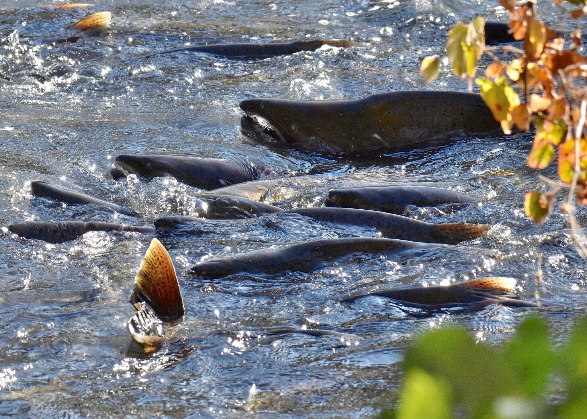
Cover Photo: Joshua Asel
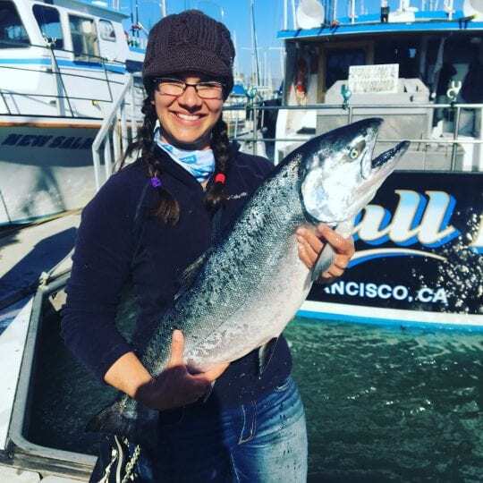
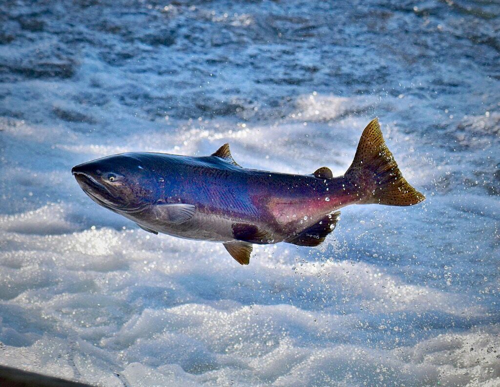
Study lead author Miranda Tilcock pioneered a technique for freshwater fish that involves peeling its eye lenses to reveal its dietary and habitat history. Here, she holds a salmon. Credit: Courtesy Miranda Tilcock/UC Davis
Fish eye lenses are onion-like spheres, rich in protein. Similar to onions, lenses are composed of individual layers that grow throughout the lifetime of a fish. Each of these layers can be peeled off one by one for isotope analysis with each layer representing a different point in time during a fish’s life. Applying stable isotope techniques with these individual layers allows researchers to better understand what and where a fish was consuming food at various points throughout its life history.
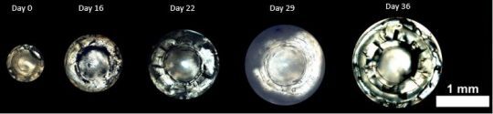
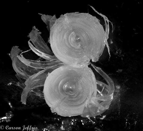
Cross section of juvenile Chinook salmon weekly lens growth on the Yolo Bypass. Time 0 represents fish from the hatchery arriving to the floodplain enclosure experiment detailed in Jeffres et al. (2020)
When we applied this technique to the single adult Chinook Salmon, we were able to see that this fish had early life history values indicative of hatchery rearing. We were then able to identify when the fish reached the estuary before moving into the ocean. Once you isolate what habitat fish are using, then you can begin to quantify it for long term success. Using Chinook Salmon as an example, we want to understand the long-term benefits that restoration and management of floodplains can provide for salmon during their juvenile life stage. We are now using this isotope tool in addition to the otoliths to reconstruct their life history of those that have returned to spawn and see what proportion spent time on the floodplain before migrating out to the ocean. Allowing us to measure the success of restoration and management actions in the Central Valley of California.
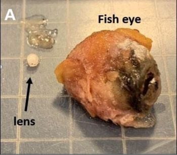
Image of an adult Chinook Salmon eye next to the lens removed from the eye.
Photo: Jane Dysert
While this technique was particularly insightful for Chinook Salmon, its applications are not limited to salmon within this region. All over the world there are migratory species in need of freshwater habitat, and many of these environments are declining in quantity and quality. Understanding which habitats are important for fish at different life stages can aid in conservation efforts.
Cross section of a lens from a tuna eye showing the multiple layers.
Photo: Jane Dysert
To learn more, listen to Miranda on NPR’s Science Friday as she tells host Ira Flatow how de-laminating fish eyeballs with tiny tweezers “is just like peeling the world’s tiniest onion.”
The study was funded by the California Department of Water Resources.
Additional study co-authors include George Whitman and Andrew Rypel, holder of the Peter B. Moyle and California Trout Endowed Chair in Cold Water Fishes at UCD, and Ted Sommer, lead scientist at the California Department of Water Resources.
Journal Reference:
- Miranda Bell‐Tilcock, Carson A. Jeffres, Andrew L. Rypel, Ted R. Sommer, Jacob V. E. Katz, George Whitman, Rachel C. Johnson. Advancing diet reconstruction in fish eye lenses. Methods in Ecology and Evolution, 2021; DOI: 10.1111/2041-210X.13543